
The proximal-transitional tail as a model for understanding vertebral diversification
Many mammal tails show a rapid morphological transition in the proximal-transitional tail, where vertebrae abruptly shift in shape, size, and articulation despite shared Hox identity. We use this transition to ask how local molecular, cellular, and tissue-level differences during embryonic development and postnatal growth give rise to diverse vertebral shape, size, and functions.
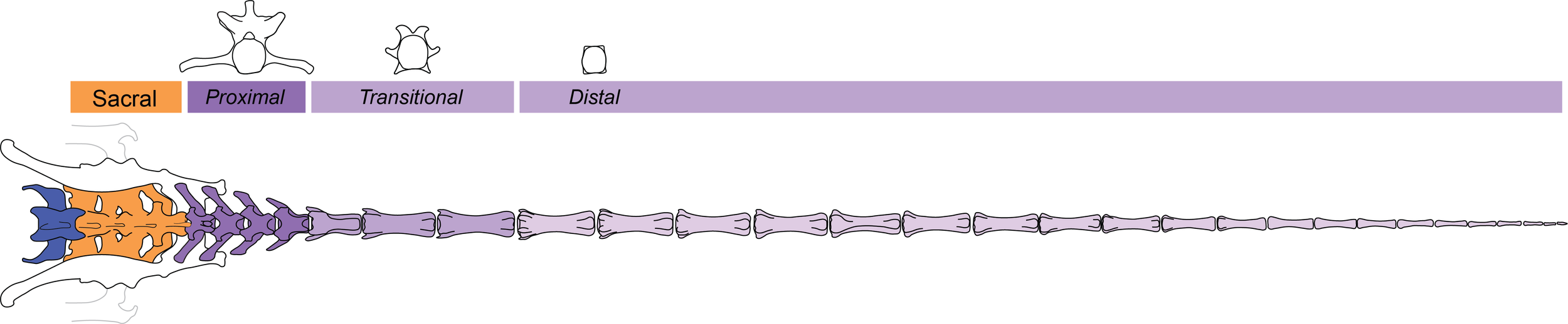
Vertebra elongation and axial proportion
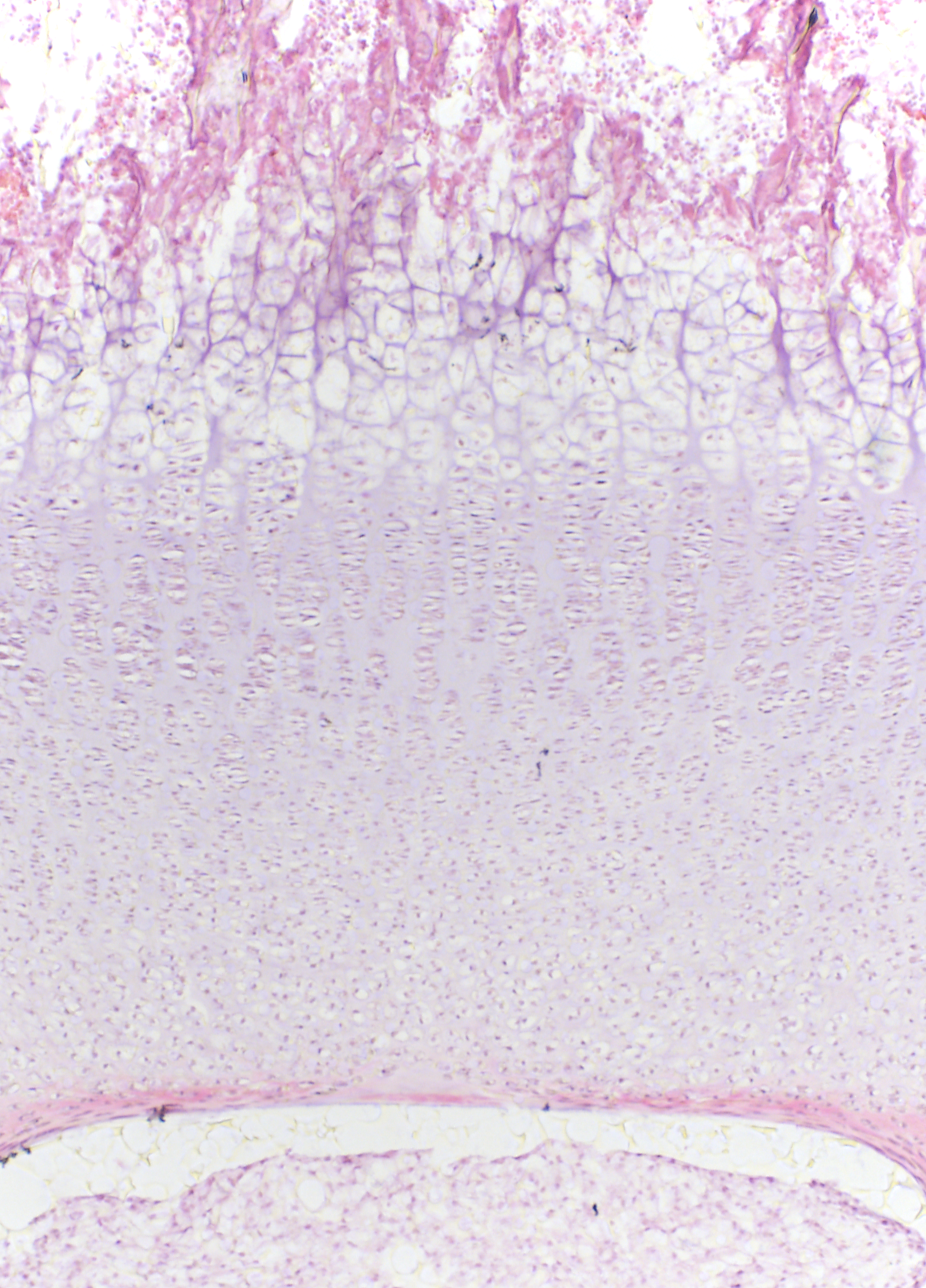
Jerboa TV6 cartilage
Hematoxylin & Eosin stain
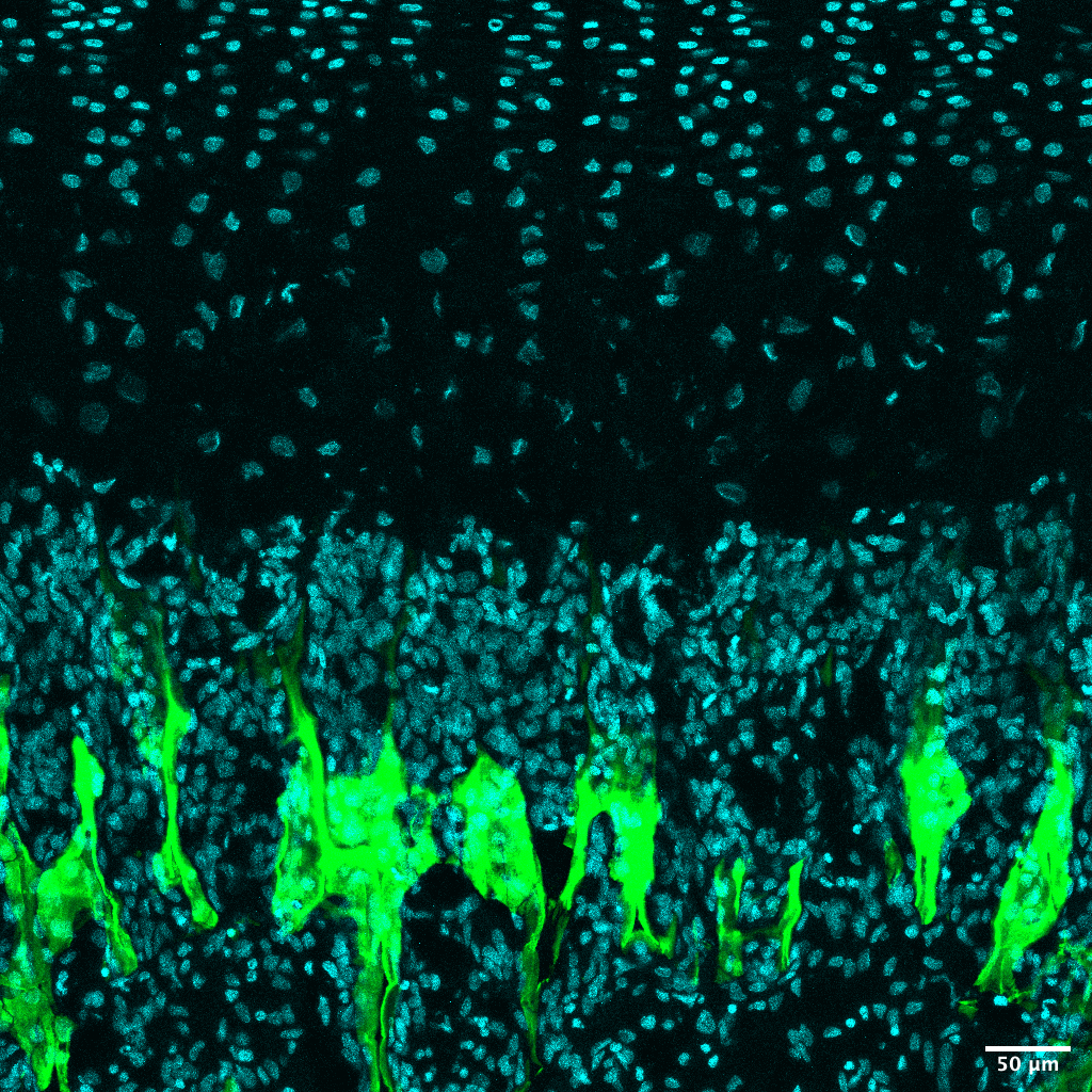
Jerboa TV6 cartilage
Calcein DAPI
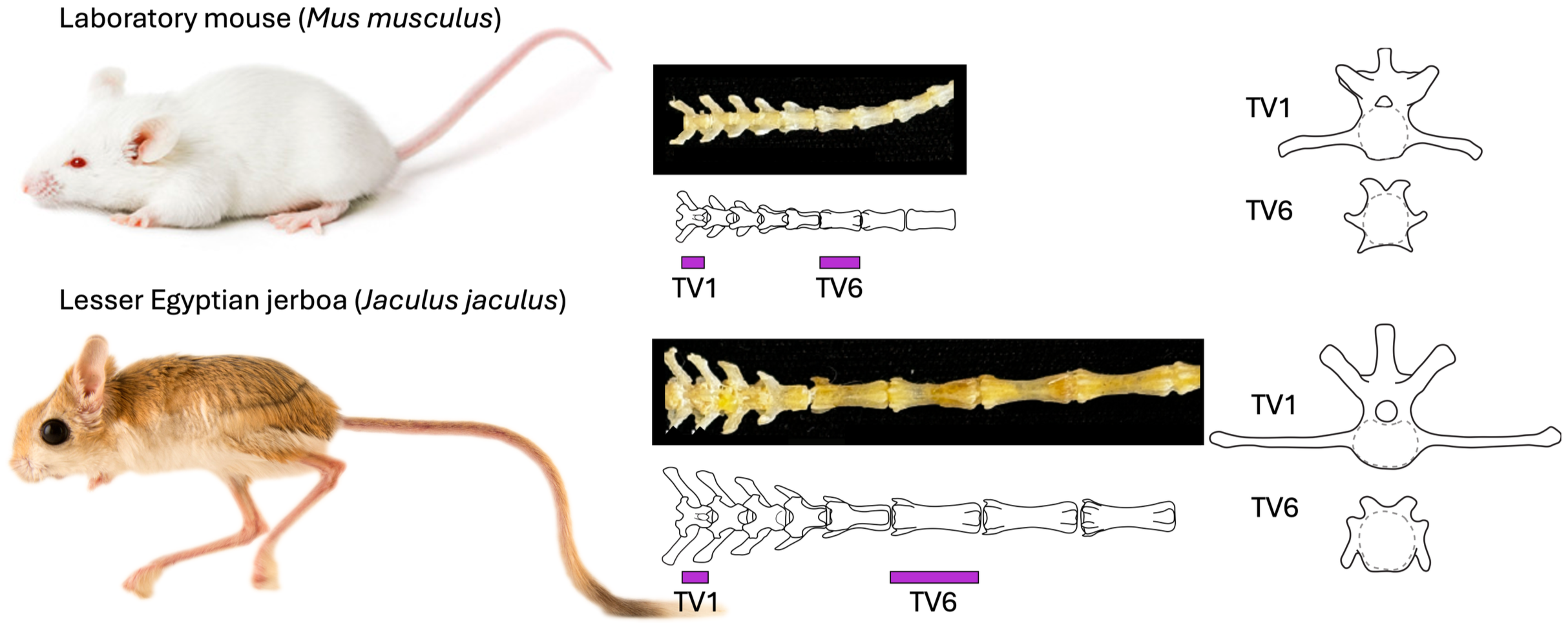
Comparative studies show that proximal tail morphology strongly correlates with tail use and total tail length, and many species exhibit a conserved “crescendo–decrescendo” pattern in which proximal vertebrae increase to a peak length and then taper distally. In collaboration with Dr. Talia Moore at the University of Michigan, we recently demonstrated that amplification of this pattern improves inertial maneuvering during locomotion (Fu 2025).
Normalized jerboa tail length is ~1.5x longer than in mice despite having fewer vertebrae, because elongation is achieved by dramatically lengthening mid-tail elements rather than increasing count. This exaggerated “crescendo–decrescendo” pattern likely contributes to their aerial agility in escape. Using jerboa and mouse, we have investigated when and how these vertebrae become disproportionate and identified both the cellular basis and candidate molecular regulators of differential elongation.
We found that larger growth plates and increased chondrocyte proliferation drive differential elongation of the vertebral body, and in the longest jerboa vertebrae this is further amplified by much larger hypertrophic chondrocytes. Intersectional transcriptomics revealed that most genes regulating vertebral proportion differ from those that control limb proportion, but a significant subset is shared in both systems. Among these is natriuretic peptide receptor C (Npr3). Loss of NPR3 in mice disproportionately elongates proximal and mid-tail vertebrae by expanding the hypertrophic zone and increasing chondrocyte size, suggesting a conserved network that modulates skeletal proportion (Weber 2025).
Vertebra morphogenesis
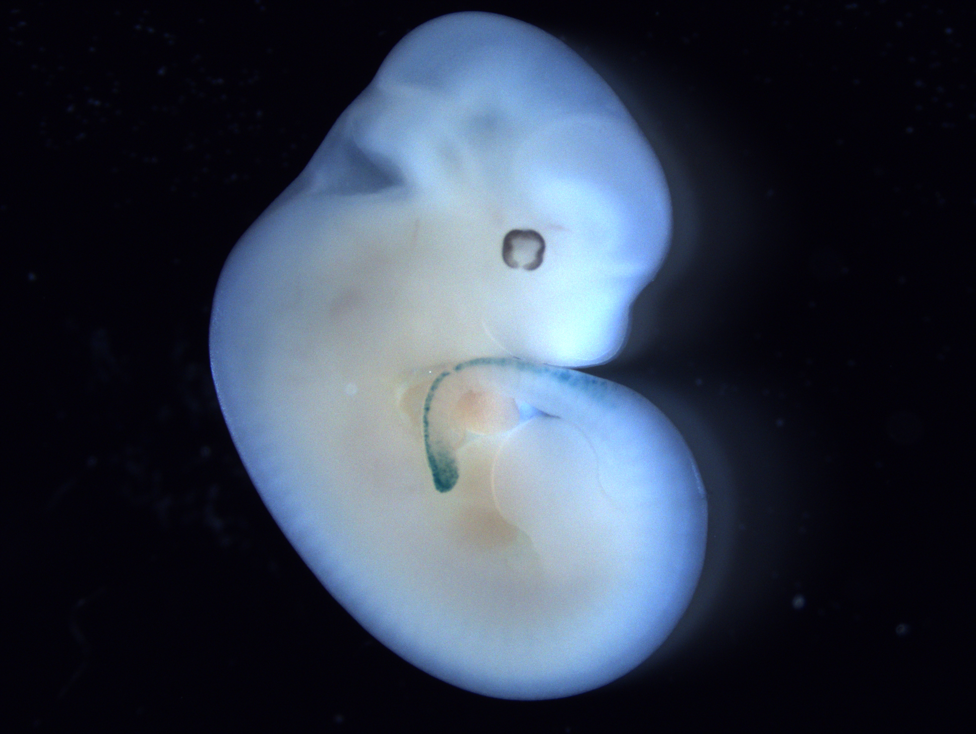
Mouse E12 Hoxb13-cre / Gt(ROSA)26Sortm1Sor
Vertebrae are serially repeated structures, yet adjacent elements can differ strikingly in both size and morphology. Mammalian vertebrae are organized into five regions defined by overlapping co-linear Hox expression: cervical (neck), thoracic (torso), lumbar (lower back), sacral (pelvis), and caudal (tail), though there can be substantial morphological variation between adjacent elements within these regions. Vertebrae arise from the somite-derived sclerotome, which is patterned into ventral, central, lateral, and dorsal domains that give rise to the vertebral body, pedicles and articular/transverse processes, ribs, and neural arch/spinous process, respectively. The proportions of these elements underlie axial skeletal function, articulation, and locomotion. These sclerotome domains are specified by inductive signals from adjacent tissues through dorsoventral and mediolateral gradients of Shh, BMPs, and Wnts. How does local modulation of signaling pathways fine-tune sclerotome specification and vertebra morphogenesis?
Evolution of diverse mammal tail morphologies and functions
The transition from pre-mammalian synapsids to mammals involved a major shift from sprawling to upright posture and a decoupling of tail and leg movement. Because an equivalent decoupling fueled adaptive radiation in theropod dinosaurs, did a release from locomotor constraint similarly allowed the mammal tail to diversify through modification of postnatal growth mechanisms? Expanding on previous studies, we have sampled tail skeletal morphology broadly across mammals to test hypotheses about the evolutionary history and drivers of tail diversification. To date, we have surveyed 245 mammal species from 119 families and 26 orders, recording sacral and tail vertebral lengths and the serial positions of traits linked to regionalization and mechanics.
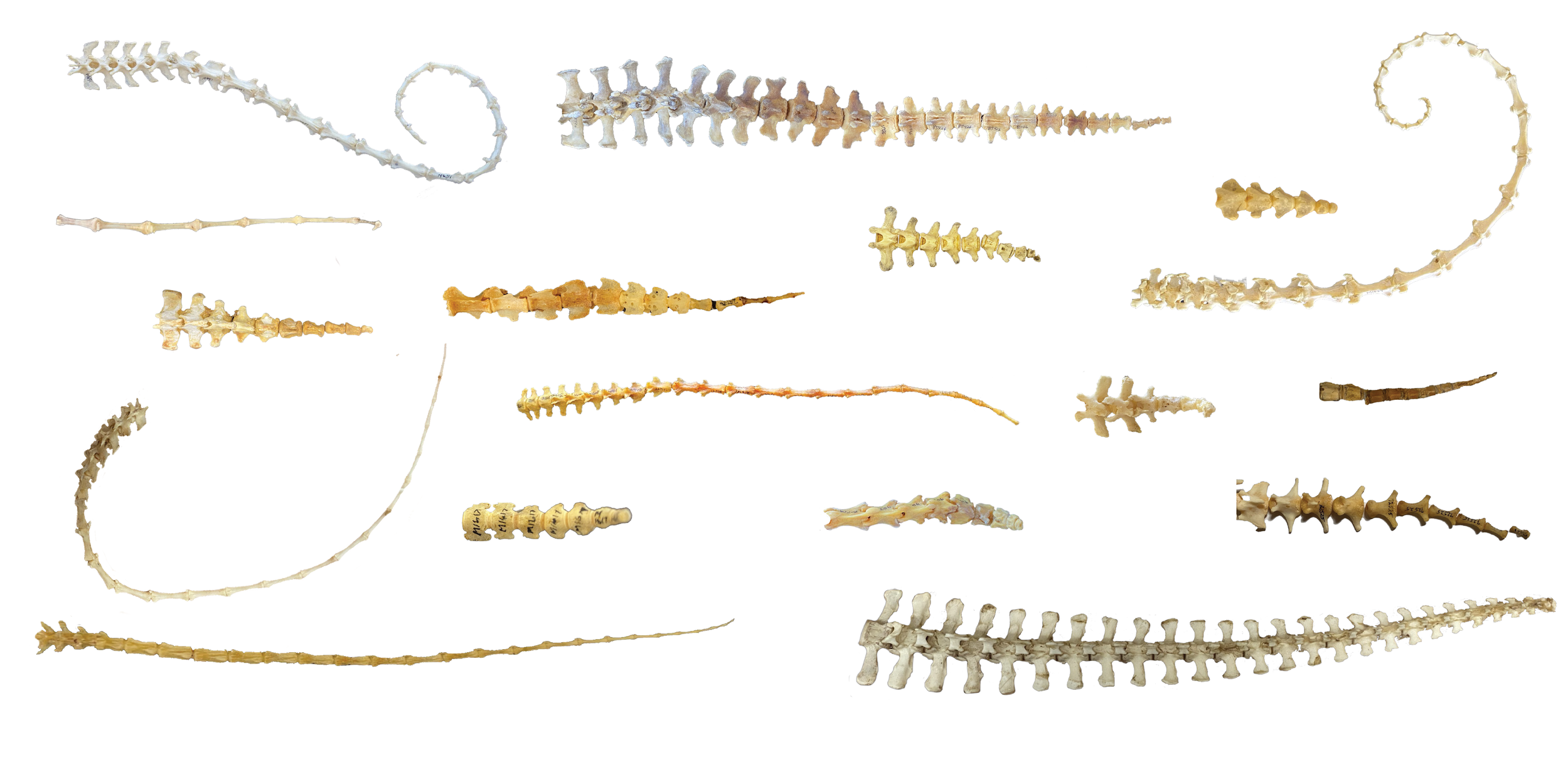
All tail skeletons pictured here are from of the San Diego Natural History museum, Natural History Museum of Los Angeles, Harvard Museum of Comparative Zoology, and University of Michigan Museum of Zoology.